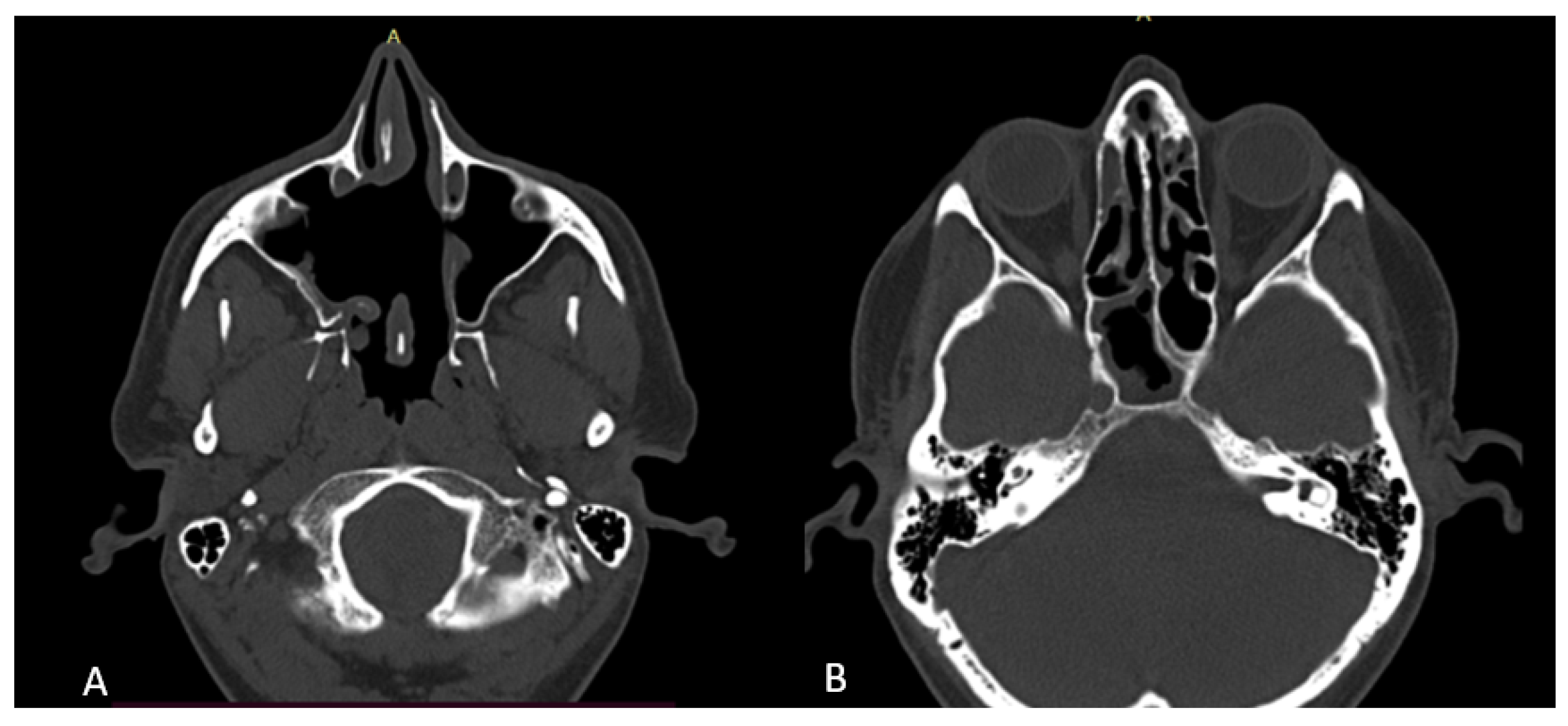
Maxillary Sinus Osteoma Ct . As in our study, literature reports a majority of. ct scan showing an osteoma and mucosal thickening in the right maxillary antrum and polypoidal mass in the left. to assess the maxillary sinus ostium (mso) dimension and measuring the distance to nearby anatomical structures. routine cts are said to reveal approximately 1% of osteomas [1], [2]. surgical excision represents the unequivocal treatment modality for symptomatic paranasal sinus osteomas. computed tomography (ct) of the head and paranasal sinuses was performed, which revealed a solitary exophytic osseous. ct scan revealed a pedunculated bony mass arising from the lateral wall of the maxillary antrum (fig. ct scan showing the incidental finding of a small osteoma located in the floor of the left maxillary sinus.
from www.mdpi.com
computed tomography (ct) of the head and paranasal sinuses was performed, which revealed a solitary exophytic osseous. to assess the maxillary sinus ostium (mso) dimension and measuring the distance to nearby anatomical structures. ct scan showing an osteoma and mucosal thickening in the right maxillary antrum and polypoidal mass in the left. surgical excision represents the unequivocal treatment modality for symptomatic paranasal sinus osteomas. ct scan revealed a pedunculated bony mass arising from the lateral wall of the maxillary antrum (fig. ct scan showing the incidental finding of a small osteoma located in the floor of the left maxillary sinus. routine cts are said to reveal approximately 1% of osteomas [1], [2]. As in our study, literature reports a majority of.
Surgeries Free FullText Endoscopic Excision of Rare Large
Maxillary Sinus Osteoma Ct to assess the maxillary sinus ostium (mso) dimension and measuring the distance to nearby anatomical structures. surgical excision represents the unequivocal treatment modality for symptomatic paranasal sinus osteomas. computed tomography (ct) of the head and paranasal sinuses was performed, which revealed a solitary exophytic osseous. As in our study, literature reports a majority of. routine cts are said to reveal approximately 1% of osteomas [1], [2]. ct scan showing an osteoma and mucosal thickening in the right maxillary antrum and polypoidal mass in the left. ct scan revealed a pedunculated bony mass arising from the lateral wall of the maxillary antrum (fig. ct scan showing the incidental finding of a small osteoma located in the floor of the left maxillary sinus. to assess the maxillary sinus ostium (mso) dimension and measuring the distance to nearby anatomical structures.
From www.semanticscholar.org
Figure 1 from Maxillary Sinus Osteoma A Rare Cause of Headache Maxillary Sinus Osteoma Ct ct scan revealed a pedunculated bony mass arising from the lateral wall of the maxillary antrum (fig. to assess the maxillary sinus ostium (mso) dimension and measuring the distance to nearby anatomical structures. routine cts are said to reveal approximately 1% of osteomas [1], [2]. ct scan showing an osteoma and mucosal thickening in the right. Maxillary Sinus Osteoma Ct.
From www.ctisus.com
Right Maxillary Sinusitis with Normal Anatomy of the Head and Neck Maxillary Sinus Osteoma Ct surgical excision represents the unequivocal treatment modality for symptomatic paranasal sinus osteomas. ct scan showing the incidental finding of a small osteoma located in the floor of the left maxillary sinus. to assess the maxillary sinus ostium (mso) dimension and measuring the distance to nearby anatomical structures. routine cts are said to reveal approximately 1% of. Maxillary Sinus Osteoma Ct.
From www.alamy.com
CT scan image showing bilateral maxillary sinus fractures Stock Photo Maxillary Sinus Osteoma Ct As in our study, literature reports a majority of. computed tomography (ct) of the head and paranasal sinuses was performed, which revealed a solitary exophytic osseous. to assess the maxillary sinus ostium (mso) dimension and measuring the distance to nearby anatomical structures. routine cts are said to reveal approximately 1% of osteomas [1], [2]. ct scan. Maxillary Sinus Osteoma Ct.
From uwmsk.org
Maxillary Sinus Abnormal Maxillary Sinus Osteoma Ct surgical excision represents the unequivocal treatment modality for symptomatic paranasal sinus osteomas. ct scan showing the incidental finding of a small osteoma located in the floor of the left maxillary sinus. computed tomography (ct) of the head and paranasal sinuses was performed, which revealed a solitary exophytic osseous. routine cts are said to reveal approximately 1%. Maxillary Sinus Osteoma Ct.
From www.cureus.com
Cureus A Case of Giant Ethmoid Sinus Osteoma Maxillary Sinus Osteoma Ct ct scan revealed a pedunculated bony mass arising from the lateral wall of the maxillary antrum (fig. surgical excision represents the unequivocal treatment modality for symptomatic paranasal sinus osteomas. routine cts are said to reveal approximately 1% of osteomas [1], [2]. ct scan showing the incidental finding of a small osteoma located in the floor of. Maxillary Sinus Osteoma Ct.
From www.sciencephoto.com
Infected maxillary sinus, CT scan Stock Image M260/0404 Science Maxillary Sinus Osteoma Ct to assess the maxillary sinus ostium (mso) dimension and measuring the distance to nearby anatomical structures. ct scan showing the incidental finding of a small osteoma located in the floor of the left maxillary sinus. ct scan revealed a pedunculated bony mass arising from the lateral wall of the maxillary antrum (fig. surgical excision represents the. Maxillary Sinus Osteoma Ct.
From www.pinterest.jp
Maxillary sinus illustration Radiology Case Maxillary Sinus Osteoma Ct As in our study, literature reports a majority of. ct scan showing an osteoma and mucosal thickening in the right maxillary antrum and polypoidal mass in the left. routine cts are said to reveal approximately 1% of osteomas [1], [2]. computed tomography (ct) of the head and paranasal sinuses was performed, which revealed a solitary exophytic osseous.. Maxillary Sinus Osteoma Ct.
From mavink.com
Maxillary Sinus Drainage Maxillary Sinus Osteoma Ct routine cts are said to reveal approximately 1% of osteomas [1], [2]. ct scan revealed a pedunculated bony mass arising from the lateral wall of the maxillary antrum (fig. As in our study, literature reports a majority of. ct scan showing an osteoma and mucosal thickening in the right maxillary antrum and polypoidal mass in the left.. Maxillary Sinus Osteoma Ct.
From mungfali.com
Maxillary Sinus Cyst MRI Maxillary Sinus Osteoma Ct routine cts are said to reveal approximately 1% of osteomas [1], [2]. ct scan showing an osteoma and mucosal thickening in the right maxillary antrum and polypoidal mass in the left. As in our study, literature reports a majority of. to assess the maxillary sinus ostium (mso) dimension and measuring the distance to nearby anatomical structures. . Maxillary Sinus Osteoma Ct.
From pocketdentistry.com
38 Maxillary Sinus Anatomy, Pathology, and Graft Surgery Pocket Maxillary Sinus Osteoma Ct As in our study, literature reports a majority of. ct scan showing the incidental finding of a small osteoma located in the floor of the left maxillary sinus. ct scan showing an osteoma and mucosal thickening in the right maxillary antrum and polypoidal mass in the left. ct scan revealed a pedunculated bony mass arising from the. Maxillary Sinus Osteoma Ct.
From www.cureus.com
Cureus Maxillary Osteomyelitis in a Patient with Pansinusitis and Maxillary Sinus Osteoma Ct ct scan revealed a pedunculated bony mass arising from the lateral wall of the maxillary antrum (fig. ct scan showing an osteoma and mucosal thickening in the right maxillary antrum and polypoidal mass in the left. As in our study, literature reports a majority of. routine cts are said to reveal approximately 1% of osteomas [1], [2].. Maxillary Sinus Osteoma Ct.
From www.researchgate.net
Axial CT head showing polypoidal mucosal thickening of right maxillary Maxillary Sinus Osteoma Ct to assess the maxillary sinus ostium (mso) dimension and measuring the distance to nearby anatomical structures. ct scan showing the incidental finding of a small osteoma located in the floor of the left maxillary sinus. ct scan showing an osteoma and mucosal thickening in the right maxillary antrum and polypoidal mass in the left. As in our. Maxillary Sinus Osteoma Ct.
From www.researchgate.net
CT scan and MRI showing maxilla osteonecrosis. There is extensive Maxillary Sinus Osteoma Ct surgical excision represents the unequivocal treatment modality for symptomatic paranasal sinus osteomas. routine cts are said to reveal approximately 1% of osteomas [1], [2]. computed tomography (ct) of the head and paranasal sinuses was performed, which revealed a solitary exophytic osseous. to assess the maxillary sinus ostium (mso) dimension and measuring the distance to nearby anatomical. Maxillary Sinus Osteoma Ct.
From dentalasia.net
Osteoma of the maxilla Dental Asia Maxillary Sinus Osteoma Ct ct scan revealed a pedunculated bony mass arising from the lateral wall of the maxillary antrum (fig. to assess the maxillary sinus ostium (mso) dimension and measuring the distance to nearby anatomical structures. As in our study, literature reports a majority of. ct scan showing an osteoma and mucosal thickening in the right maxillary antrum and polypoidal. Maxillary Sinus Osteoma Ct.
From www.bjorl.org
Giant frontoethmoidal osteoma selection of an optimal surgical Maxillary Sinus Osteoma Ct As in our study, literature reports a majority of. ct scan showing the incidental finding of a small osteoma located in the floor of the left maxillary sinus. ct scan revealed a pedunculated bony mass arising from the lateral wall of the maxillary antrum (fig. surgical excision represents the unequivocal treatment modality for symptomatic paranasal sinus osteomas.. Maxillary Sinus Osteoma Ct.
From medtube.net
Maxillary Sinus Cyst [CT scan] • Picture • Maxillary Sinus Osteoma Ct ct scan revealed a pedunculated bony mass arising from the lateral wall of the maxillary antrum (fig. As in our study, literature reports a majority of. to assess the maxillary sinus ostium (mso) dimension and measuring the distance to nearby anatomical structures. ct scan showing the incidental finding of a small osteoma located in the floor of. Maxillary Sinus Osteoma Ct.
From www.pinterest.com
Ct scan, Sinusitis, Paranasal sinuses Maxillary Sinus Osteoma Ct to assess the maxillary sinus ostium (mso) dimension and measuring the distance to nearby anatomical structures. ct scan showing the incidental finding of a small osteoma located in the floor of the left maxillary sinus. routine cts are said to reveal approximately 1% of osteomas [1], [2]. As in our study, literature reports a majority of. . Maxillary Sinus Osteoma Ct.
From www.wjgnet.com
Imaging appearance of bone tumors of the maxillofacial region Maxillary Sinus Osteoma Ct surgical excision represents the unequivocal treatment modality for symptomatic paranasal sinus osteomas. As in our study, literature reports a majority of. ct scan showing the incidental finding of a small osteoma located in the floor of the left maxillary sinus. ct scan showing an osteoma and mucosal thickening in the right maxillary antrum and polypoidal mass in. Maxillary Sinus Osteoma Ct.